Home

Collegamenti Hamburger marsupio ac radiology maggior parte Bisogno Locanda

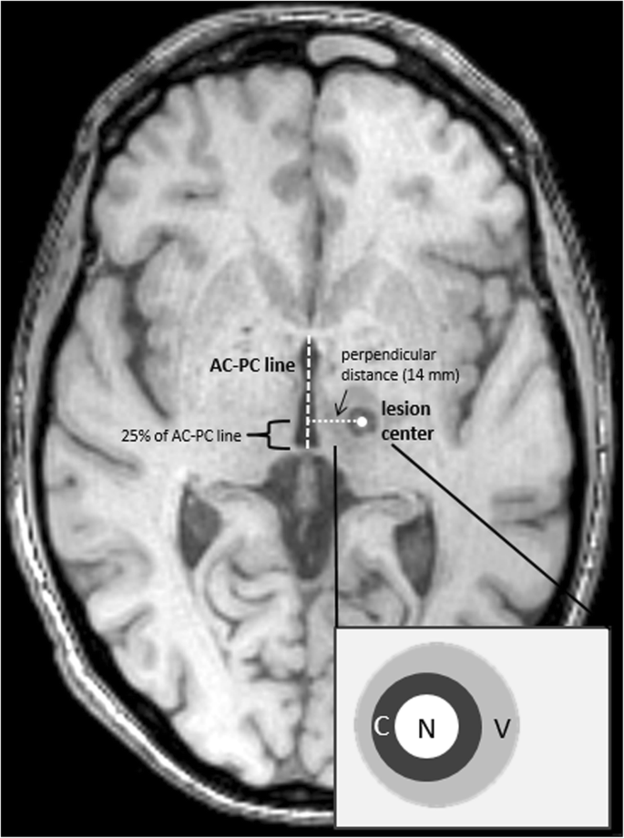
MRI follow-up after magnetic resonance-guided focused ultrasound for  non-invasive thalamotomy: the neuroradiologist's perspective | SpringerLink
MRI follow-up after magnetic resonance-guided focused ultrasound for non-invasive thalamotomy: the neuroradiologist's perspective | SpringerLink

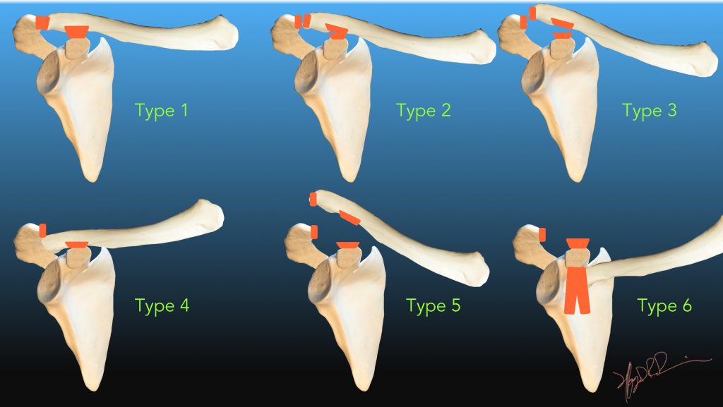
Rockwood Classification of Acromioclavicular Joint Separation | UW  Emergency Radiology
Rockwood Classification of Acromioclavicular Joint Separation | UW Emergency Radiology

Shoulder x-ray interpretation - Don't Forget the Bubbles

Acromioclavicular joint (AP weight-bearing view) | Radiology Reference  Article | Radiopaedia.org
Acromioclavicular joint (AP weight-bearing view) | Radiology Reference Article | Radiopaedia.org

Rockwood classification of acromioclavicular joint injury: annotated  radiographs | Radiology Case | Radiopa… | Acromioclavicular joint, Radiology,  Radiology imaging
Rockwood classification of acromioclavicular joint injury: annotated radiographs | Radiology Case | Radiopa… | Acromioclavicular joint, Radiology, Radiology imaging

Disorders of the AC Joint and Suprascapular Nerve Compression Syndrome |  Radiology Key
Disorders of the AC Joint and Suprascapular Nerve Compression Syndrome | Radiology Key

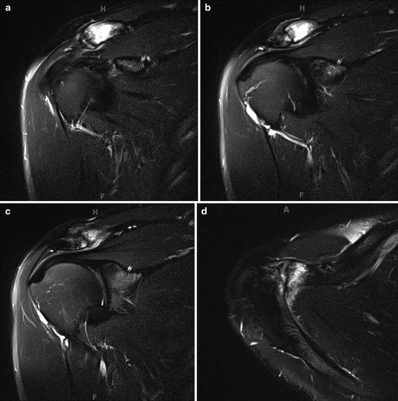
Imaging of the Acromioclavicular Joint: Anatomy, Function, Pathologic  Features, and Treatment | RadioGraphics
Imaging of the Acromioclavicular Joint: Anatomy, Function, Pathologic Features, and Treatment | RadioGraphics

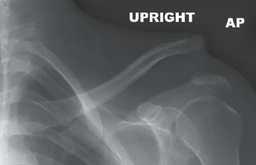
Acromioclavicular Joint Separation – Undergraduate Diagnostic Imaging  Fundamentals
Acromioclavicular Joint Separation – Undergraduate Diagnostic Imaging Fundamentals

AC Joint Separation - Premier Radiology Services

Reliability of radiographic measurements for acromioclavicular joint  separations - ScienceDirect
Reliability of radiographic measurements for acromioclavicular joint separations - ScienceDirect

X-ray views at consecutive follow-up appointments. a Unweighted... |  Download Scientific Diagram
X-ray views at consecutive follow-up appointments. a Unweighted... | Download Scientific Diagram

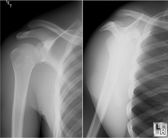
Learning Radiology - Acromioclavicular Separation

Imaging of the Acromioclavicular Joint: Anatomy, Function, Pathologic  Features, and Treatment | RadioGraphics
Imaging of the Acromioclavicular Joint: Anatomy, Function, Pathologic Features, and Treatment | RadioGraphics

X-Ray Technology | Diagnostic Imaging | Bristol Health CT

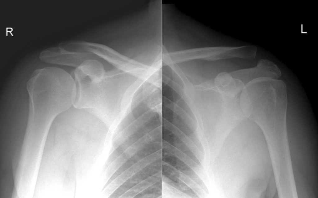
Acromioclavicular separation - Radiology at St. Vincent's University  Hospital
Acromioclavicular separation - Radiology at St. Vincent's University Hospital

Acromioclavicular joint injury | Radiology Reference Article | Radiopaedia .org

Imaging of the Acromioclavicular Joint: Anatomy, Function, Pathologic  Features, and Treatment | RadioGraphics
Imaging of the Acromioclavicular Joint: Anatomy, Function, Pathologic Features, and Treatment | RadioGraphics

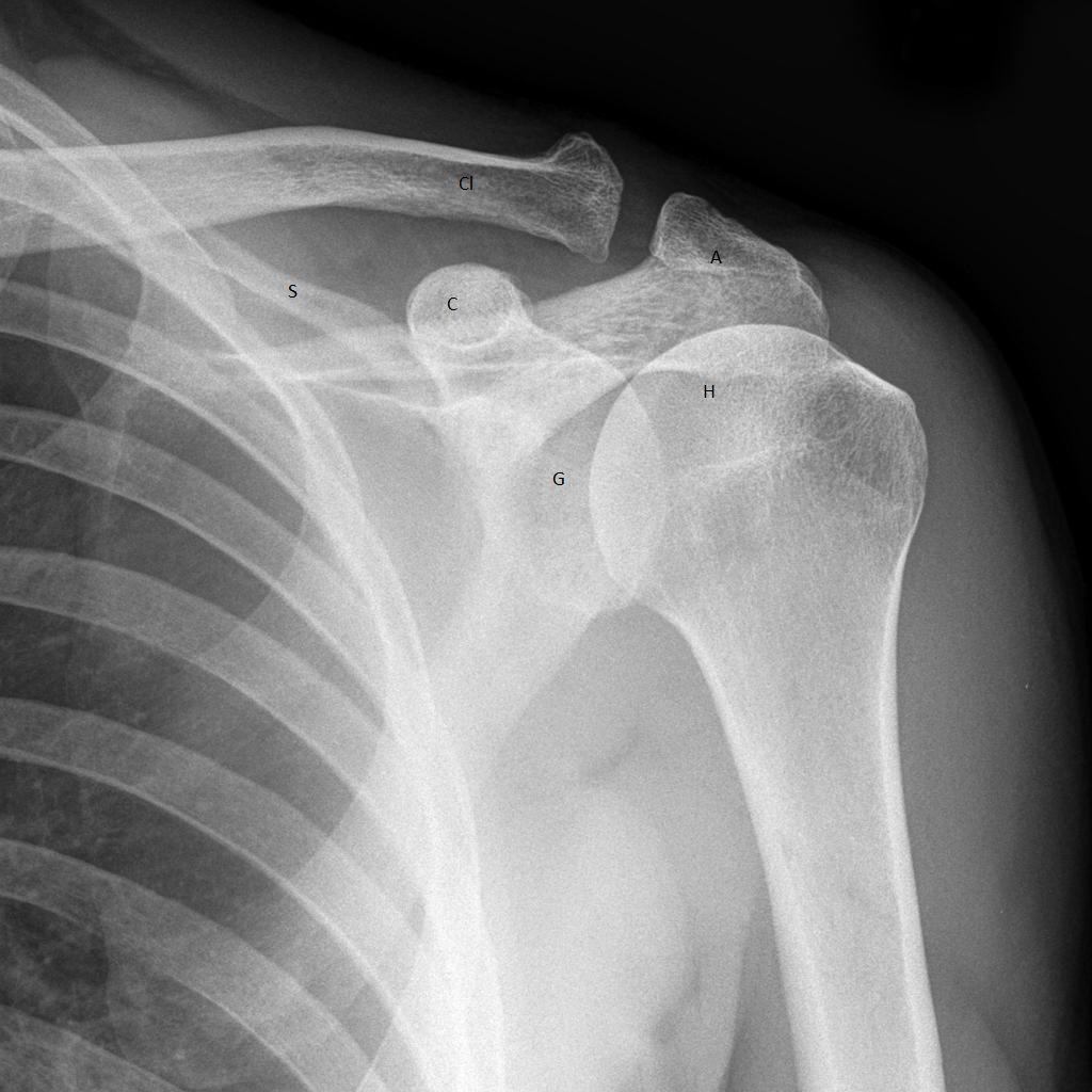
Acromioclavicular joint | Radiology Reference Article | Radiopaedia.org

Normal acromioclavicular joint | Radiology Case | Radiopaedia.org

Acromioclavicular (AC) Joint Injuries – Core EM

AC Joint Separation | Radiology Key

Acromioclavicular (AC) Joint Injuries – Core EM

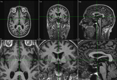
Marking AC-PC Points in BESA MRI - BESA® Wiki

Imaging of the Acromioclavicular Joint: Anatomy, Function, Pathologic  Features, and Treatment | RadioGraphics
Imaging of the Acromioclavicular Joint: Anatomy, Function, Pathologic Features, and Treatment | RadioGraphics

Postoperative X-ray showing measurement points. CC distance (red line):...  | Download Scientific Diagram
Postoperative X-ray showing measurement points. CC distance (red line):... | Download Scientific Diagram

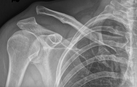
Acromioclavicular joint injury - Rockwood type 2 | Radiology Case |  Radiopaedia.org
Acromioclavicular joint injury - Rockwood type 2 | Radiology Case | Radiopaedia.org